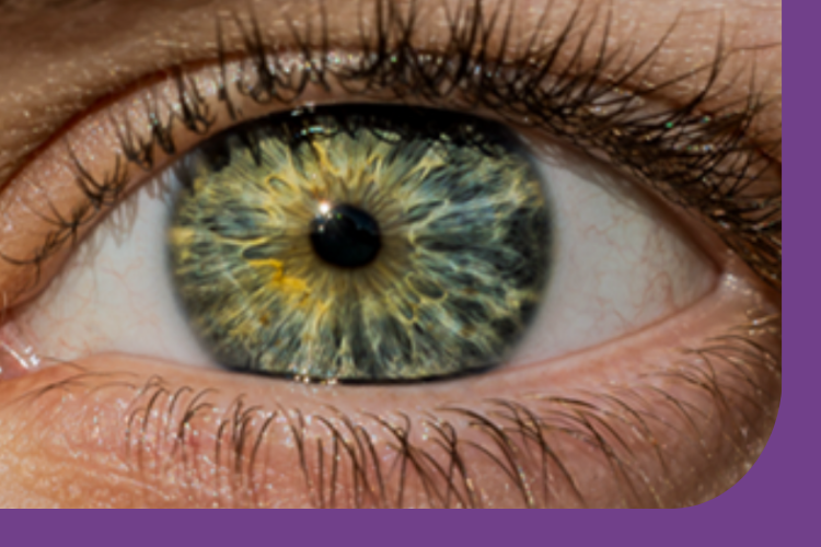
Not much is known about how changes in the retina, the light-sensitive layer at the back of the eye, may reflect what is happening in the brain of people living with motor neuron disease (MND) or dementia.
Using a quick and easily accessible eye scan called OCT, we can take detailed images of the retina - which offers a unique “window” into brain health. Because the retina and brain are closely connected, studying the retina may help us understand how nerve cells are damaged in these conditions.
Why is it important?
Currently, there are few reliable ways to measure or monitor nerve damage in people living with MND or dementia. This makes it difficult to track how these conditions progress and how well treatments are working. OCT offers a simple and non-invasive way to explore subtle retinal changes that could appear before symptoms become more noticeable.
Most research so far has focused on the brain itself. We know much less about how MND and dementia affect the retina, even though these changes could provide valuable clues about disease mechanisms. The RET–MND study will therefore:
-
Use OCT scans to look for patterns of retinal change in people living with MND or dementia
-
Explore how these changes relate to symptoms
-
Identify early retinal features that could help with earlier diagnosis and monitoring
What could we learn?
RET–MND could reveal how the eye mirrors change in the brain, helping detect neurodegenerative conditions earlier, monitor disease progression more accurately, and guide the development of new treatments. Ultimately, this research could transform how we study and care for people living with MND or dementia—using the eye as a powerful, available tool to understand these conditions.
Image by kind permission of Miracle Ozzoude


